×
Select Your Country
 International

×
Select Your Country
 International


The flow of blood from the heart to the rest of the organs is controlled by the aortic valve. Transcatheter Aortic Valve Implant (TAVI) is a minimally invasive procedure that is performed for replacing the diseased or damaged aortic valve with a new artificial one with the help of a narrow tube called the catheter. This tube is placed inside a large blood vessel in the chest or groin.
Why Manipal?
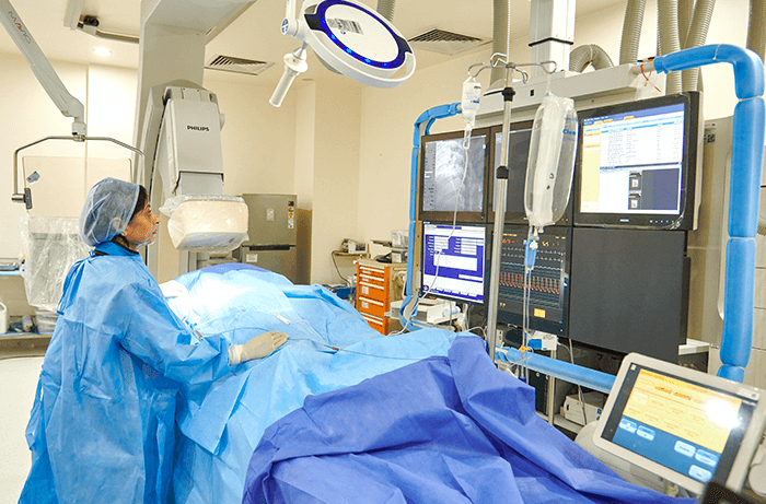
At Manipal Hospitals, we are equipped with a world-class cardiac catheterization lab, which is furnished with advanced high precision technology to undertake complex surgeries, such as the TAVI. Our extensively trained team of cardiac surgeons, cardiac anaesthetists, and interventional cardiologists specialize in cardiac ultrasound imaging, which helps them perform this procedure with ease.
Solutions
TAVI is a minimally invasive catheterisation procedure that is performed for fixing a defective aortic valve. This valve generally opens when the heart pumps blood to the rest of the body. The inability of this valve to open and close properly puts excessive strain on the heart, thus causing blackouts, breathlessness, or dizziness.
Risks
Although the TAVI is the standard procedure for aortic valve malfunction, it can cause some complications in patients if not conducted properly. In some instances, patients with valve replacements may be highly vulnerable to infections of the heart valve and the surrounding tissue known as endocarditis. In some cases, excessive bleeding occurs in geriatric people during the procedure.
Success of TAVI
For aortic valve replacement, TAVI has become the best alternative to open-heart surgeries. Moreover, the popularity of this procedure is surging sharply globally owing to its safety and durability.
Infection Control Protocol
At Manipal Hospitals, we consider the safety and comfort of the patients to be of utmost priority. Post the TAVI procedure, the patient will be monitored in our surgical intensive care unit for a minimum of 24 hours to prevent the occurrence of any side effects or infections.
Left Atrial Appendage and Closure
The left atrial appendage (LAA) is basically an ear-shaped and small sac in the muscle wall of the left atrium, which lies in the top left chamber of the heart. A healthy heart contracts during each heartbeat and the blood in the LAA and the left atrium is pushed towards the left ventricle, which is the bottom left chamber of the heart. In a person who has an irregular heartbeat, the electrical impulses that control the heartbeat are chaotic and fast, which does not give the atria the time to contract or push blood into the ventricles. This can cause the formation of blood clots in the atria and LAA. The pumping of these blood clots out of the heart can cause a stroke. If the patient is at risk of developing blood clots in the LAA or left atrium, the doctor may recommend a procedure to close the LAA, which will, in turn, mitigate the chances of strokes and the need to consume blood-thinning medications.
Solutions of LAA
Implants are the latest treatment options available for LAA. At Manipal Hospitals, one of the best heart hospitals in India we have highly experienced cardiologists who implant the WATCHMAN device through the patient’s skin in the Cath Lab under general anaesthesia. The procedure involves the insertion of a catheter sheath into a vein located near the groin, which is then guided across the septum – the muscular wall that divides the left and rides sides of the heart – and towards the LAA’s opening. The device is implanted at the opening of the LAA, which closes the LAA completely, thus preventing the formation of clots.