Kidney Stones Treatment for Cameroon Patients Diagnosis to Recovery
Reviewed by: Nephrology Department
Posted on Jul 14, 2026
12 Min Read
Synopsis
What Are Kidney Stones (Nephrolithiasis)?
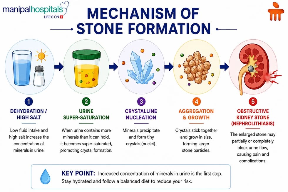
Kidney stones are solid masses formed when minerals and salts present in urine become concentrated and crystallize inside the kidneys. These crystals gradually stick together, forming stones that vary in size from tiny grains of sand to larger stones capable of blocking urine flow.
Several types of kidney stones exist, including:
- Calcium oxalate stones: The most common type, formed from calcium and oxalate.
- Uric acid stones: Often associated with dehydration, high-protein diets, or gout.
- Struvite stones: Usually develop following recurrent urinary tract infections.
- Cystine stones: A rare form caused by an inherited metabolic disorder.
It is clinically vital to distinguish between true kidney stones and urinary bladder calculi. While both are manifestations of urolithiasis, kidney stones form in the upper urinary tract (renal pelvis and calyces). Urinary bladder calculi, conversely, develop primarily in the lower urinary tract, often due to incomplete bladder emptying caused by conditions like benign prostatic hyperplasia (BPH) or urethral strictures, though they can occasionally form when a small renal stone migrates downward and grows within the bladder.
Symptoms of Kidney Stones
The clinical presentation of nephrolithiasis varies based on the stone's size, surface texture, and location within the urinary collecting system. A stone remaining stationary within the renal pelvis may cause vague, dull flank aches or remain entirely asymptomatic. However, when a stone moves into the narrow ureter, it triggers acute, severe symptoms:
- Severe Flank Pain (Renal Colic): A sharp, excruciating pain located in the flank (the side and back, just below the ribs). This pain typically fluctuates in intense waves as the muscular walls of the ureter spasm to force the stone downward.
- Radiating Pain: As the stone descends, the pain trajectory shifts, radiating downward into the lower abdomen, pelvis, groin, and occasionally the labia or testicles.
- Dysuria: A painful, burning sensation during urination, which occurs when a stone approaches or lodges near the ureterovesical junction (where the ureter meets the bladder).
- Haematuria: Blood in the urine, causing it to appear pink, red, or tea-coloured, due to structural friction against the delicate mucosal lining of the urinary tract.
- Changes in Urine Quality: Turbid (cloudy) or foul-smelling urine, indicating the presence of pyuria (white blood cells) or concurrent bacterial colonisation.
- Urinary Urgency and Frequency: A persistent, increased urge to urinate, even when the bladder holds minimal volume.
- Systemic Gastrointestinal Symptoms: Nausea and vomiting, triggered by shared nerve pathways between the kidneys and the gastrointestinal tract (the celiac plexus)
Environmental and Lifestyle Context in Cameroon
In Cameroon, distinct environmental and regional factors heavily influence these physiological risks. The tropical climate, characterised by prolonged dry seasons and high temperatures—particularly in the Sudano-Sahelian northern regions (such as Garoua and Maroua)—induces rapid sweating and high insensible fluid loss. Without adequate intake of clean, potable water, urine quickly becomes super-saturated. Furthermore, local dietary habits, including a heavy reliance on dried or highly salted fish and meats for preservation, alongside regional variations in mineral content within local water sources, can elevate urinary sodium and calcium levels, necessitating hyper-vigilant hydration strategies across all regions.
When Should You See a Doctor?
While renal colic is intensely painful, certain red-flag symptoms signal a medical emergency, such as an obstructed, infected kidney.
Patients must seek immediate emergency medical evaluation if they experience:
- Fever and chills (indicative of a dangerous, ascending systemic infection).
- An absolute inability to pass urine (complete urinary tract obstruction).
- Persistent, uncontrolled vomiting that prevents the retention of oral fluids or medications.
- Intractable pain unresponsive to standard oral analgesics.
- Gross, heavy hematuria featuring large blood clots
How Are Kidney Stones Diagnosed?
Accurate diagnosis is the first step toward effective therapy for kidney stones. Identifying the stone's size, location, composition, and any associated complications enables doctors to develop a personalised treatment plan that offers the best possible outcomes.
At Manipal Hospitals Global, international patients benefit from a comprehensive diagnostic approach supported by advanced imaging technologies, modern laboratory services, and multidisciplinary expertise. From initial evaluation to treatment planning, experienced urologists, nephrologists, radiologists, and laboratory specialists work together to ensure timely and accurate diagnosis.
Medical History and Physical Examination
The diagnostic process begins with a detailed consultation. Your doctor will review your symptoms, previous history of kidney stones, dietary habits, daily fluid intake, family history, medications, and any underlying medical conditions. A physical examination helps identify tenderness in the abdomen or back and assesses your overall health to guide further investigations.
Urine Tests
Urinalysis is an essential part of diagnosing kidney stones and helps detect the following:
- Blood in the urine (hematuria)
- Signs of urinary tract infection
- Crystals associated with different stone types
- Abnormal urine composition
For patients with recurrent nephrolithiasis, a 24-hour urine collection may be recommended to evaluate metabolic abnormalities and identify factors contributing to repeated stone formation.
Blood Tests
Blood investigations provide important information about kidney health and possible causes of stone formation. These tests typically assess:
- Kidney function (creatinine and urea)
- Calcium levels
- Uric acid levels
- Signs of infection or inflammation
The results help specialists determine whether an underlying metabolic or medical condition is contributing to kidney stone development.
Advanced Imaging Studies
Imaging is crucial for confirming the diagnosis, determining the stone's exact size and location, and selecting the most appropriate treatment approach.
Ultrasound (echography) is often the first imaging investigation. It is a safe, non-invasive, and radiation-free technique that effectively detects many kidney stones and evaluates the kidneys for swelling or urinary obstruction.
A CT scan (computed tomography) is considered the gold standard for diagnosing kidney stones. It provides highly detailed images that accurately identify the number, size, and precise location of stones while helping rule out other causes of abdominal or flank pain.
In selected cases, X-rays may also be used to monitor certain types of stones during treatment or follow-up.
Minimally Invasive Kidney Stones Treatment Options
| Procedure | Indication & Stone Size | How It Works (Mechanism) | Recovery & Hospital Stay |
| ESWL (Shock Wave Lithotripsy) | Small-to-moderate stones (< 1.5–2 cm) in the kidney or upper ureter. | External acoustic shock waves fracture the stone into fine, sand-like fragments without incisions. | Outpatient (0 days). Rapid recovery; resume normal activities within 24–48 hours. |
| URS (Ureteroscopy with Laser) | Stones located anywhere throughout the ureter or renal pelvis. | An endoscopic scope is passed through the urethra; a precise laser breaks the stone. A temporary stent is often placed. | 0–1 day hospital stay. Return to normal activities within 3–5 days. |
| PCNL (Percutaneous Nephrolithotomy) | Large, complex, or staghorn stones (> 2 cm) in the kidney. | The procedure was performed under general anaesthesia through a small 1 cm incision in the back to directly fragment and extract the stones. | 2–4 days hospital stay. Full physical recovery takes 2–4 weeks. |
| Open / Robotic Surgery | Complex anatomical issues or severe structural damage; exceptionally rare. | Surgical incisions are made to physically remove the stone when minimally invasive options fail. | Extended hospital stay. Longer recovery timeline depending on complexity. |
Comprehensive Diagnostic Care at Manipal Hospitals Global
At Manipal Hospitals Global, patients have access to advanced diagnostic facilities designed to support accurate and timely kidney stone evaluation. Our specialists combine clinical expertise with modern imaging and laboratory diagnostics to determine the most effective treatment strategy. This comprehensive approach enables personalised care, faster diagnosis, and seamless treatment planning for both domestic and international patients, ensuring every patient receives world-class care from consultation through recovery.
Preventing Future Kidney Stones
People who have experienced one kidney stone have a higher chance of developing another. Fortunately, several lifestyle changes can significantly reduce this risk.
Preventive measures include:
- Drink enough clean water throughout the day to maintain clear or pale-yellow urine.
- Limit excessive salt intake, as sodium increases calcium excretion into urine.
- Consume a balanced diet rich in fruits and vegetables.
- Avoid excessive consumption of sugary beverages.
- Moderate animal protein intake.
- Maintain a healthy body weight.
- Follow medical advice regarding calcium intake rather than eliminating calcium completely.
- Manage underlying health conditions such as gout or recurrent urinary tract infections.
- Attend regular follow-up appointments if you have previously been diagnosed with nephrolithiasis.
Individuals who develop recurrent stones may benefit from specialised metabolic evaluation and dietary counselling to identify specific risk factors.
When Should You Seek Immediate Medical Attention?
Although many kidney stones pass without surgery, some situations require urgent evaluation.
Seek immediate medical care if you experience:
- Severe pain that cannot be controlled with medication
- Fever or chills alongside urinary symptoms
- Persistent vomiting
- Inability to urinate
- Heavy bleeding in the urine
- Symptoms that worsen despite treatment
Prompt intervention can prevent kidney damage, severe infection, and other potentially life-threatening complications.
Conclusion
Kidney stones are a common and highly painful condition, but they are completely treatable through modern medicine. Early clinical diagnosis using reliable imaging modalities like ultrasound or a non-contrast CT scan allows for highly personalised and effective care. Today's advanced therapy for kidney stones offers excellent options tailored to every patient's needs, from conservative medical management for small stones to state-of-the-art, minimally invasive procedures for larger obstructions.
By combining timely medical intervention with long-term preventive strategies, such as consistent hydration, balanced nutrition, and regular medical follow-ups, you can successfully eliminate stones, protect your renal health, and enjoy a lasting, pain-free recovery.
If you are experiencing persistent flank pain, blood in your urine, or symptoms of kidney stones, consult the urology specialists at Manipal Hospitals Global for advanced diagnosis and minimally invasive treatment. Our international patient team can assist with appointments, travel planning, and personalised treatment guidance.
Share this Article


Not sure what you’re looking for?
Not sure what you’re looking for?
Get in touch with our representatives for
help!
Get in touch with our
representatives for help!